The use of positron emission tomography (PET) for disease diagnosis and repeated monitoring of the effectiveness of medical treatments is on the rise, with the number of required scans increasing at an estimated rate of about 11% per year. Keeping up with this demand may pose challenges to radiology departments, generating a need for PET scanners that are faster to operate, less costly to purchase and require fewer scan-related personnel resources.
With this aim, researchers at Ghent University in Belgium are developing a novel patient-centred upright imaging device for total body PET – the walk-through TB-PET. Their proposed TB-PET design, which is visually similar to an airport security scanner, is expected to be over three times cheaper than a cylindrical long axial field-of-view (LAFOV) system and reduce radiographer/technologist time by more than half of that required for standard axial field-of-view (SAFOV) PET scanners.
The walk-through device will employ monolithic detectors with depth-of-interaction (DOI) capabilities and high intrinsic spatial resolution to produce high-quality PET images in sub-minute scans.
Writing in the European Journal of Nuclear Medicine and Molecular Imaging, the researchers describe the dual flat-panel design of the walk-through TB-PET and present performance comparisons – in terms of component costs, system sensitivity, patient throughput and required dose per patient – with a SAFOV PET scanner (Biograph Vision 600) and a LAFOV scanner (Vision Quadra). They note that the comparison presumes the walk-through scanner will be used for efficient routine clinical PET imaging, and is therefore based on scans of only the torso and head.
Principal investigator Stefaan Vandenberghe and co-researchers have proposed a new design concept for PET imaging that relies on two opposing flat-panel detectors that can be brought as close as reasonably possible to the patient (standing upright between them) to increase both sensitivity and spatial resolution.
The team selected monolithic detectors that offer two to three times higher spatial resolution than the pixelated detectors used in today’s clinical PET systems. The ability of monolithic detectors to encode DOI information provides a uniform spatial resolution of just below 2 mm over the whole field-of-view. These detectors have an expected coincidence timing resolution between 200 and 400 ps.

The walk-through TB-PET scanner is composed of two flat panels, each about 70 cm wide and 106 cm high, with a 50 cm gap between them. Each panel consists of a 14 × 20 array of monolithic bismuth germanate (BGO) detector blocks, 50 × 50 × 16 mm in size, read out by an array of 6 x 6 mm silicon photomultipliers. The team derived the scanner size and flat panel dimensions using body measurements obtained from PET/CT images of 40 randomly selected patients from CHU de Liège.
During a scan, the patient will stand between the two flat-panel detectors. Crucially, this walk-through design removes the need for time-consuming positioning of the patient on and off the bed. Another advantage is the system’s small footprint, which only requires about 2–6 m2 of dedicated space, a fraction of the installation spaces required for today’s PET imaging suites (35–40 m2). Scanner cooling requirements should also be less.
The workflow comparison generated impressive findings. Vandenberghe and colleagues estimated that the walk-through TB-PET could scan up to 87 patients during an eight-hour shift, in comparison with 53–60 patients for the LAFOV scanner and 28 for the SAFOV system.
The researchers estimated the cost of their proposed scanner based on monolithic BGO or lutetium-yttrium oxyorthosilicate (LYSO) scintillators, as well as the cost of the silicon photomultipliers, which represent the two main expenses in a PET scanner. They determined that the component cost for a BGO-based walk-through TB-PET is 3.3 times lower than that of a LAFOV system with a 106 cm axial field-of-view, and just 20% higher than that of a SAFOV scanner.
Innovative devices ramp the resolution of PET imaging
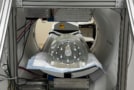
After constructing a mock-up of the scanner, the researchers discovered that the addition of handlebars could significantly reduce the motion of a patient in a standing position. They are also using the mock-up to determine whether breath-hold is feasible with 30 s acquisitions, and plan to test techniques for motion estimation and correction.
Other future plans include constructing a system by assembling the detectors into modules, building a patient platform with automated patient height adjustment of the flat panels, and integrating motion detection. Ultimately, the researchers aim to integrate the walk-through scanner with a standing CT to combine molecular imaging with high-resolution anatomical imaging. They also hope to further reduce the PET and CT dose using advanced deep-learning-based noise reduction methods.
- SEO Powered Content & PR Distribution. Get Amplified Today.
- PlatoData.Network Vertical Generative Ai. Empower Yourself. Access Here.
- PlatoAiStream. Web3 Intelligence. Knowledge Amplified. Access Here.
- PlatoESG. Automotive / EVs, Carbon, CleanTech, Energy, Environment, Solar, Waste Management. Access Here.
- PlatoHealth. Biotech and Clinical Trials Intelligence. Access Here.
- ChartPrime. Elevate your Trading Game with ChartPrime. Access Here.
- BlockOffsets. Modernizing Environmental Offset Ownership. Access Here.
- Source: https://physicsworld.com/a/walk-through-pet-scanner-made-for-high-throughput-imaging-at-lower-cost/