An ultrahigh-resolution 7 tesla (T) MRI scanner seven years in development can generate functional brain images with 10 times better spatial resolution than current 7 T scanners, and over 180 times more detail than conventional 3 T systems. As reported in Nature Methods, this improved spatial resolution reveals functional MRI (fMRI) features as small as 0.35 mm, compared with the typical 2–3 mm of standard 3 T fMRI.
“The scanner was designed for ultrahigh spatial resolution to identify neuronal activity at different depths in the cerebral cortex, allowing brain circuitry to be studied by differentiating activity in different cortical cell layers,” explains project director David Feinberg, of the Helen Wills Neuroscience Institute at UC Berkeley. Such high-resolution fMRI is not possible using current 7 T scanners, because the signal decays before it can be recorded by the scanner’s hardware systems.
Feinberg explains that the improved resolution will help researchers examine neuronal circuits in different parts of the brain and track signals propagating from one area of the cortex to another while a person is thinking and reasoning. Pinpointing the fMRI activity to a specific depth in the cortex reveals activity in feedforward and feedback circuitry localized at different cortical depths, enabling neuroscientists to determine the direction of information movement throughout the brain.
As well as improving fMRI, the new NexGen 7 T scanner also offers higher spatial resolution in diffusion, physiological and structural MR imaging. The team plans to use it to study underlying changes in brain circuitry in various brain disorders, including degenerative diseases, schizophrenia and development disorders such as autism.
Scanner design
The NexGen 7 T scanner is based on a commercial MAGNETOM Terra 7 T scanner from Siemens Healthineers. To increase the spatial resolution, Siemens scientists – in collaboration with researchers at Harvard-MIT Health Sciences and Technology and UC Berkeley – developed the Impulse head-only gradient coil, which incorporates an additional third layer of wire windings and a powerful cooling system.
Shielded gradient coils in a standard MRI scanner include two layers of conductive wiring. The inner layer generates linear magnetic fields for spatial encoding of images and the outer layer minimizes eddy currents in the surrounding superconducting magnet by cancelling the external magnetic fields. The addition of a third layer of wire winding in the Impulse gradient coil provides the additional degrees of freedom needed to reduce peripheral nerve stimulation, as well as optimize the gradient field linearity, mechanical resonances and torque.
The Impulse gradient coil achieves much faster gradient switching (900 T/m/s) and higher maximum gradient amplitude (200 mT/m) than standard 7 T whole-body gradient coils. As such, it can achieve an order of magnitude greater performance than current standard 7 T systems, and about five times the performance of an existing head-only gradient coil operating at 7 T.
The NexGen 7 T scanner is also the first to incorporate a 128-channel receiver system. The scanner currently includes two RF receiver–transmit coil arrays: a 64-channel receiver array coil and a 96-channel coil, with 4 cm loop diameters. These larger receiver arrays with smaller coil loops achieve higher signal in the cortex and roughly 30% improvement in signal-to-noise ratio compared with a standard 32-channel receiver array coil.
Other system modifications required to optimize the new scanner included adapting the acquisition computer to support the large data set size acquired with 64-, 96- and 128-channel receiver coil arrays and the larger matrix sizes used for high-resolution imaging. This included increasing the memory capacity of the reconstruction computer to superfast memory modules and expanding the disk space for raw k-space data on the measurement and reconstruction system. The team also designed a custom interface connector to accommodate 128 receive channels.
Ultrahigh-field MRI reveals brain changes in migraine sufferers
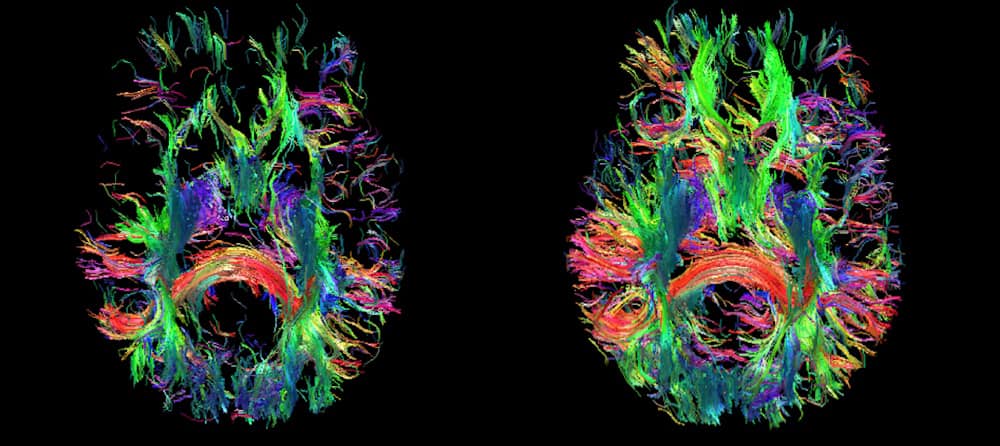
To date, the researchers have imaged over 100 healthy volunteers on the NexGen 7 T scanner with no adverse effects. Studies included evaluating multi-echo echo-planar imaging, cerebral blood volume contrast imaging, diffusion imaging, gradient echo imaging and MR angiography. They note that future studies with co-registration of structural and functional data in a single participant, combining high angular resolution of fibre tracks and laminar fMRI of the whole brain should be possible.
Many technical challenges remain before super-resolution MRI scanners can be used in routine clinical brain imaging. High cost is another factor. But the team believes that “the numerous innovations developed and incorporated into the NexGen 7 T scanner will make diverse human neuroscience studies at ultrahigh resolution routinely possible, including functional imaging of cortical layer and columnar organization”.
- The NextGen 7 T scanner was developed by a multi-institutional team of researchers at a cost of $22 million, with an initial $13.4 million research grant from the NIH’s Brain Research through Advancing Innovative Neurotechnologies (BRAIN) initiative. Academics and scientists at Siemens Healthcare, RF coil designer MR CoilTech and R&D company Advanced MRI Technologies (AMRIT) were major contributors to the scanner’s development.
- SEO Powered Content & PR Distribution. Get Amplified Today.
- PlatoData.Network Vertical Generative Ai. Empower Yourself. Access Here.
- PlatoAiStream. Web3 Intelligence. Knowledge Amplified. Access Here.
- PlatoESG. Carbon, CleanTech, Energy, Environment, Solar, Waste Management. Access Here.
- PlatoHealth. Biotech and Clinical Trials Intelligence. Access Here.
- Source: https://physicsworld.com/a/next-generation-7-t-scanner-ramps-the-resolution-of-brain-mr-imaging/